Introduction to anatomy and physiology/ Introduction to Human Body as a Whole
Table of Contents
Define the term Anatomy & Physiology (A&P)
Human Body:
The human body is a highly technical and sophisticated machine. It operates as a single entity, but is made up of a number of systems
Two branches of science, anatomy and physiology, provide the foundation for
understanding the body’s parts and functions
Anatomy:
Anatomy is the study of the structure of the body and the physical relationship between its constituent parts.
Physiology:
Physiology is the study of how the body systems work. Physiology is subdivided into:
- Viral Physiology
- Bacterial Physiology
- Cellular Physiology
- Plant Physiology
- Human Physiology
Human Physiology:
Human physiology explains the specific characteristics and mechanisms of the human body that make it a living being.
Structure (Anatomy) and functions (Physiology) are so closely related:
Understand the relationship between A&P:
Anatomy
- Tells the structure of the organ
- Tells physical relationship between body parts.
Physiology
- Tells the function of the organ
- Tells the way in which organs integrated activities maintain life and health of the individual.
Define level of organization of the body:
There are six level of organization of body which helps us understand the anatomy and
physiology.
Chemical Level:

- Chemical level is the most fundamental organizational level of body.
- In these, Atoms combines to form molecules.
Cellular Level:
- Molecules combine to form cells.
- Cells are the smallest and basic unit of living organisms.
- Trillion of cells present in human body.
- Cells are too small to be seen with the naked eye.
Tissue Level:

Cells with similar structures and functions are found together
and forming tissue.
There are just four basic types of tissue in your body:
- Epithelial tissue
- Connective tissue
- Muscular tissue
- Nervous tissue
Organ Level:
- Organs are made up of a number of different types of tissue and have
- evolved to carry out a specific function.
- Examples of organs are the stomach, skin, bones, heart, liver, lungs, and brain
System Level:
Systems consist of a number of organs that together contribute to one
or more survival needs of the body.
Organism Level/ Human body:
Human body composed of many organs systems that work together
to perform the function of an independent individual.
Define anatomical position:
Descriptions of any region or part of the human body assume
that it is in a specific stance called the anatomical position.
Some anatomical positions are discussed below:
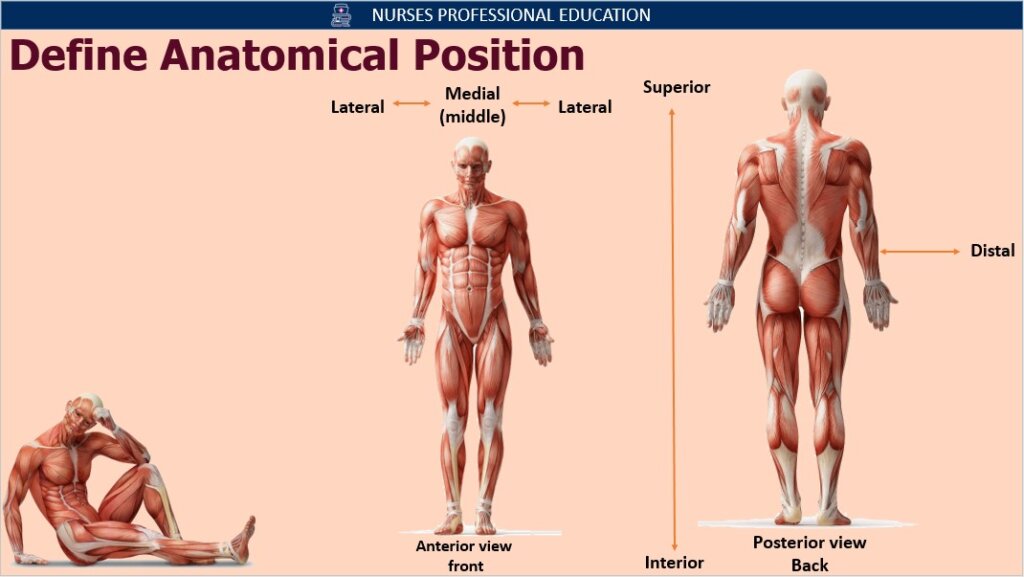
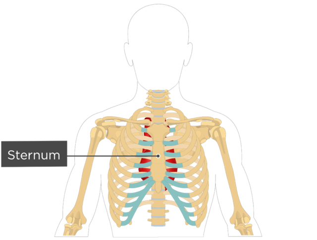
Anterior: Nearer to or at the front of the body. The sternum (breastbone) is anterior to the heart
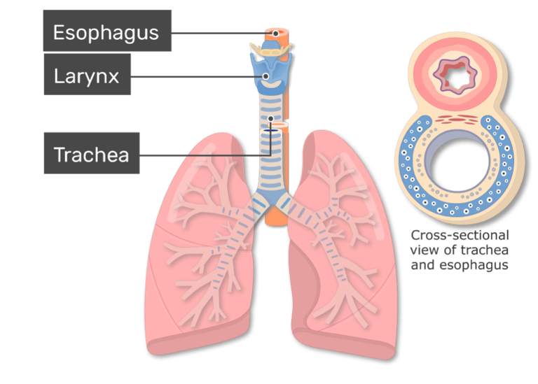
Posterior: Nearer to or at the back of the body. The esophagus is posterior to the trachea (windpipe)
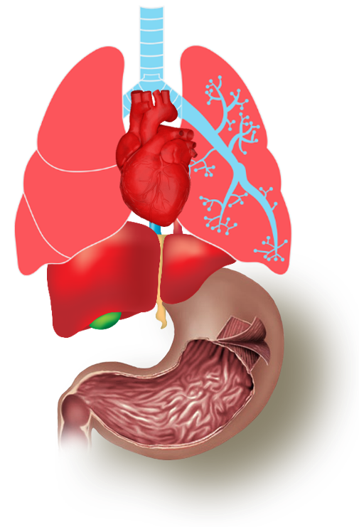
Superior: Toward the head, or the upper part of a structure. The heart is superior to the liver
Inferior: Away from the head, or the lower part of a structure. E.g. The stomach is inferior to the lung
Describe the various body planes:
Planes are used to divide the body and its parts. Some major planes of body are given below:
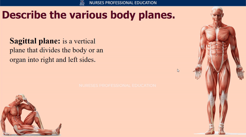
Sagittal Plane : The sagittal plane is a vertical plane that divides the body or an organ into right and left sides.
Frontal or Coronal plane : A frontal or coronal plane divides the body or an organ into anterior (front) and posterior (back) portions.
Transverse plane: A transverse plane divides the body or an organ into superior (upper) and inferior (lower) portions. Transverse plane is also known as cross-sectional and horizontal plane.
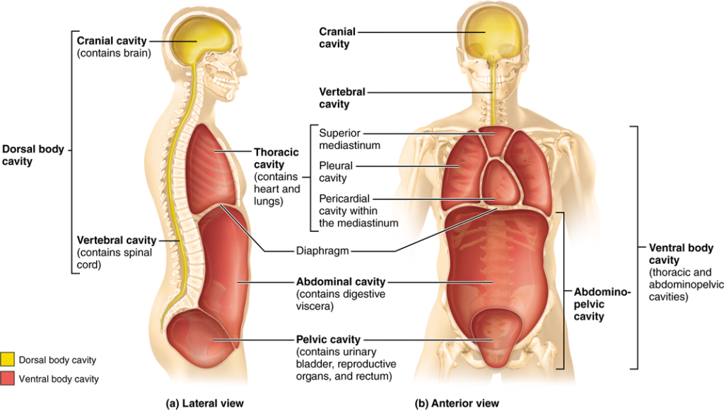
Discuss body cavities and list the organs lying within each cavity:
Body cavities are spaces within the body that help protect, separate, and support internal organs. Body cavities are given below:
Cranial Cavity: Cranial cavity formed by cranial bones and contains the brain.
Vertebral Cavity: Formed by the vertebral column and contains the spinal cord and the beginnings of spinal nerves.
Thoracic Cavity: Thoracic cavity is situated in the upper part of the trunk. The thoracic cavity is formed by the ribs,
muscle of the chest, sternum, and thoracic portion of the vertebral column. Thoracic cavity
contains pleural and pericardial cavities and mediastinum.
Pericardial Cavity: PERI= Around, CARDIAL= Heart Surrounds the heart, the serous membrane of the pericardial cavity is called pericardium.
Pleural Cavity: Each surrounds a lung; the serous membrane of each pleural cavity is called pleura.
Abdominal Cavity: This is the largest body cavity and is oval shape contains stomach, spleen, liver,
gallbladder, small intestine, and most of the large intestine. The serous membrane of the abdominal cavity is the peritoneum.
Pelvic Cavity: The pelvic cavity is roughly funnel shaped, and extends from the lower end of the abdominal cavity. The pelvic cavity contains the urinary bladder, portions of the large intestine, and internal organs of reproduction.
Oral Cavity: the Oral cavity contains the tongue and teeth.
Orbital Cavity: Orbital cavities contain the eyeball.
Middle Ear Cavity: Middle ear cavity contains small bones in the middle ear.
Synovial Cavity: Synovial cavities are found in freely movable joints and contain synovial fluid.
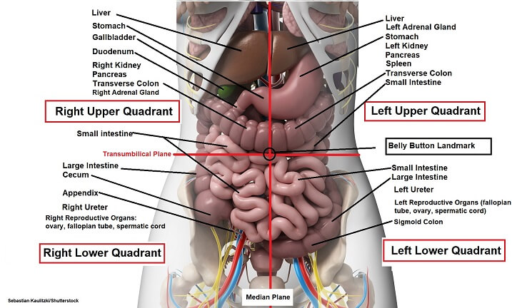
Identify abdominal pelvic region & quadrant:
The abdominopelvic cavity is subdivided into four quadrants and nine regions.
Abdominal Quadrants:
The quadrant designation is used to locate the site of pain, tumor, or some other
abnormality. There are four quadrants in abdominopelvic cavity.
LEFT UPPER QUADRANT (LUQ):
The left upper quadrant is the location of the left portion of the liver, the larger portion of
the stomach, the pancreas, left kidney, spleen, portions of the transverse and descending
colon, and parts of the small intestine. Pain in this region is associated with rotation of the intestine and colon.
RIGHT UPPER QUADRANT (RUQ):
The right upper quadrant contains the right portion of the liver, gallbladder, right kidney, a
small portion of the stomach, portions of the ascending and transverse colon, and parts of
the small intestine. Pain in this region is associated with infection and inflammation in the gallbladder and liver or peptic ulcers in the stomach.
LEFT LOWER QUADRANT (LLQ):
The left lower quadrant houses the majority of the small intestine, some large
intestine, the left female reproductive organs, and the left ureter. Pain in this region is generally associated with colitis (inflammation of the large intestine) as well as pelvic inflammatory disease and ovarian cysts in females.
RIGHT LOWER QUADRANT (RLQ):
In the right lower quadrant sits the cecum, appendix, part of the small intestines, the right
female reproductive organs, and the right ureter. Pain in this region is most commonly associated with appendicitis.
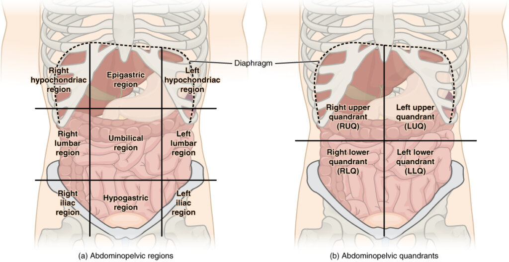
Abdominopelvic Regions:
The nine regions of the abdominopelvic cavity are smaller than the four abdominopelvic
quadrants. The nine regions division is more widely used for anatomical studies The regions of abdominopelvic cavity are:
RIGHT HYPOCHONDRIAC REGION:
The right hypochondriac region contains the right portion of the liver, the gallbladder, the
right kidney, and parts of the small intestine.
EPIGASTRIC REGION:
The Epigastric (above stomach) region contains the majority of the stomach, part of the
liver, part of the pancreas, part of the duodenum, part of the spleen, and the adrenal
glands. This region pushes out when the diaphragm contracts during breathing.
LEFT HYPOCHONDRIAC REGION:
The left hypochondriac region contains part of the spleen, the left kidney, part of the
stomach, the pancreas, and parts of the colon.
RIGHT LUMBAR REGION:
The right lumbar region consists of the gallbladder, the left kidney, part of the liver, and the
ascending colon.
UMBILICAL REGION:
The umbilical region contains the umbilicus (navel), and many parts of the small intestine,
such as part of the duodenum, the jejunum, and the ileum. It also contains the transverse
colon (the section between the ascending and descending colons) and the bottom portions
of both the left and right kidney.
LEFT LUMBAR REGION:
The left lumbar region consists of the descending colon, the left kidney, and part of the
spleen.
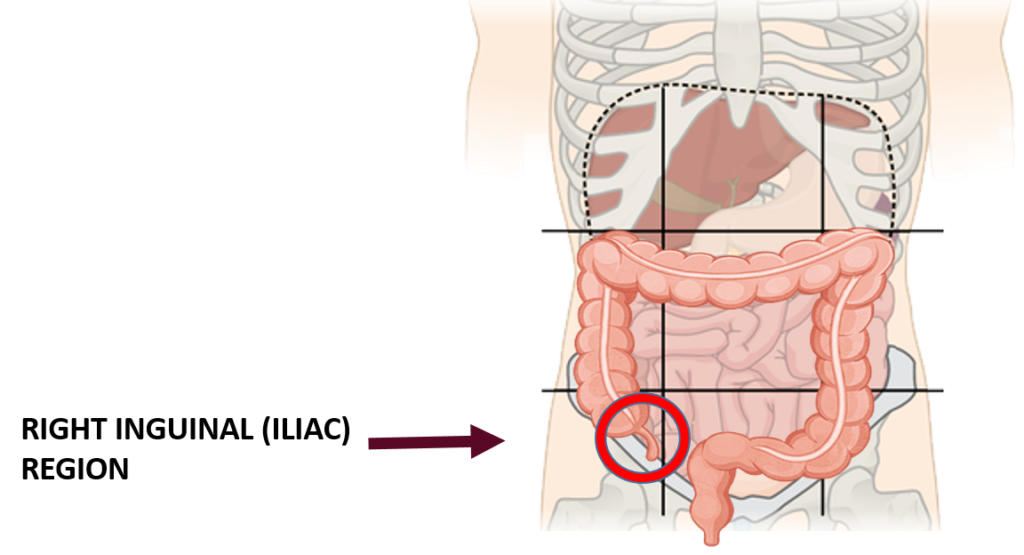
RIGHT INGUINAL / ILIAC REGION:
The right iliac region contains the appendix, cecum, and the right iliac fossa. It is also
commonly referred to as the right inguinal region. Pain in this area is generally associated
with appendicitis.
HYPOGASTRIC/PUBIC REGION:
The hypo-gastric region (below the stomach) contains the organs around the pubic bone.
These include the bladder, part of the sigmoid colon, the anus, and many organs of the
reproductive system, such as the uterus and ovaries in females and the prostate in males.
LEFT INGUINAL/ ILIAC REGION:
The left iliac region contains part of the descending colon, the sigmoid colon, and the left
iliac fossa. It is also commonly called the left inguinal region.
Briefly discuss the importance of abdomino pelvic quadrants and regions:
The human abdomen is divided into quadrants and regions by anatomists and physicians for the purposes of study, diagnosis, and treatment.
The division into four quadrants allows the localization of pain and tenderness, scars,
lumps, and other items of interest, narrowing in on which organs and tissues may be
involved.
For example, if someone having pain in their right iliac region, there must be chances that it’s because of appendicitis which needed to be checked immediately.
Name the quadrants of the abdomen and name the organ in them?
There are four abdomen quadrants in the human body.
Right upper quadrant
Right lower quadrant
Left upper quadrant
Left lower quadrant.
Organs in the right upper quadrants.
Liver
Gallbladder
Duodenum
Head of the pancreas
R adrenal glands
R kidney
Hepatic flexure of the colon
A portion of the ascending and transverse colon
Pylorus
Organs in the right lower quadrants.
The lower pole of the right kidney
Cecum
Appendix
Ascending colon
Rt ovary and fallopian tube
Rt spermatic cord
Rt ureter
Organs in the left upper quadrants.
Left lobe of the liver
Sleep
Stomach
Body of pancreas
Left adrenal gland
Left kidney
Spleenic flexure of the colon
Parts of the transverse and descending colon
Organs in the left lower quadrants.
The lower pole of the left kidney
Sigmoid colon
Left ovary and fallopian tube
Left spermatic cord
Left ureter
Define anatomy and name its different branches.
Anatomy :
Anatomy is the study of internal and external structures and physical relationships between body parts.
Anatomy is derived from the Greek word “ana” means “up” and “tomy” means cutting.
There are two main types of anatomy.
Microscopic anatomy: it is the study of internal and external structure that can be seen with the help of a microscope.
Cytology: it is the study of structures of the cell.
Histology: it is the study of the structure of the tissues
Embryology: it deals with the structure that emerges from the time of fertilization.
Macroscopic anatomy: it is the study of structure that can be seen with the naked eye; it’s also known as gross anatomy.
It is subdivided into:
Regional anatomy: structure of specific regions I.e. cephalic region, cervical region, pelvic region etc.
Systemic anatomy: it is the study of structures of major organ systems. I.e. respiratory system, digestive system.
Surface anatomy: anatomical landmarks on the surface body through visualization and palpation.
References:
- Guyton, A. C. (2001). Medical Physiology (10th ed) Washington: Kirokawa.
- Ross, & Wilson. (2000) Anatomy & Physiology in Health & Illness. Edinburgh: Churchill 8th Edition.
- Tortora, G. J. (2000). Principles of Human Anatomy and Physiology (3rd ed). New York: Happer & Row.
Introduction to the Body as a Whole Quiz:
Introduction to the Body as a Whole Complete PDF Notes :
Unit 1 Introduction to Human Body as a Whole PPT
PURCHASE
- Editing rights
- Without watermark.
- For purchasing, contact us via email (nursesprofessional.edu@gmail.com)
FREE DOWNLOAD
Enjoy free Product
- No editing rights
- With Watermark
Informative & creative 😍
Thank you so much.